
Keratoconus, often abbreviated KC, is a condition that affects the cornea, the transparent, light-focusing front surface of the eye. Over time, the cornea thins and eventually bulges into a cone shape. Since the cornea should be round, the cone-like shape causes visual alterations and difficulties, including blurred vision, glare, and light sensitivity. Let’s briefly examine how vision functions to comprehend how keratoconus may affect your vision.
Light that enters the eye travels to the brain and forms the images we recognise as vision. The cornea focuses light entering the eye, which must pass through the pupil, the inner ocular lens, and the retina, which transmits a signal to the brain. Together, the cornea and lens focus the light onto the retina. As the cornea is the entry site for light, a misshapen cornea cannot focus the light on the correct path to complete the process. This can make even routine daily duties extremely difficult! In and of itself, keratoconus is not considered a disability; however, severe vision impairment qualifies as a disability.
Causes and Symptoms
Despite many years of research, the cause of keratoconus is still a mystery. Doctors have identified genetic factors (10% of KC patients have a parent with the disease). Eye allergies and excessive eye irritation have been identified through research as risk factors. Certain connective tissue disorders can also affect corneal tissue and contribute to a keratoconus diagnosis. Many adolescents are diagnosed with schizophrenia, while others do not exhibit symptoms until adulthood. Keratoconus can affect either one or both eyes and more than fifty per cent of patients are male.
Before their diagnosis, many people have the extremely frustrating experience of being unable to see as well with their spectacles or contact lenses as they once did. This typically prompts their eye doctor to conduct additional tests or refer them to a specialist. Other indicators a person may exhibit include:
- hazy vision
- glare from lighting
- diminished night vision
- photosensitive sensitivity
- eye irritation (the sensation that something is in the eye).
- Migraines
Diagnosis and Treatment
To diagnose keratoconus, the cornea must be measured for its curvature, thickness, and overall shape. Your physician or their staff will conduct these procedures and take additional measurements. Pachymetry, which measures corneal thickness, and Keratometry, which measures corneal shape, are tests that are administered routinely. Both are non-invasive and quick. A corneal map will be created on a computer using topography or tomography. These tests have advanced significantly and can now detect even the earliest indicators of keratoconus. Your doctor will examine your vision and evaluate the overall health of your eyes’ internal and external structures. Finally, if your keratoconus is being treated with contact lenses, your doctor will position trial lenses on your eyes to determine the optimal fit and vision.
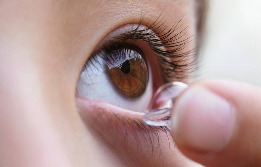
Vision loss due to keratoconus cannot be restored, but the disease can be effectively managed. Treatments for KC have also vastly advanced over the past few years. Not too long ago, the most common treatments were corneal RGP lenses (the small ones) and special flexible lenses. In recent years, scleral lenses have significantly revived and impacted the keratoconus community.
Scleral lenses are among the most common treatments. Scleral lenses alleviate the symptoms of keratoconus by creating a new, spherical surface that focuses light on the remainder of the eye. They do not land or linger on the cornea. Instead, they land on the sclera, the white part of the eye, just outside the cornea. This enhances comfort and enables your doctor to include the precise and intricate measurements necessary to create custom lenses.