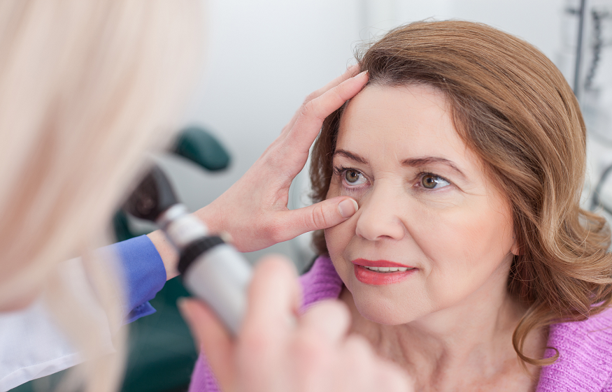
Pupillary evaluation is a key component of the neurological exam. It involves the measurement of the pupil’s size and response to light, known as pupil reactivity. Pupil reactivity is essential for assessing neurological function and diagnosing neurological conditions.
This article will discuss the importance of measuring pupil reactivity for neurological assessment, how to measure pupil reactivity accurately, and its interpretation.
What is Pupil Reactivity?
Pupil reactivity refers to the responsiveness of the pupil to light. The size of the pupil changes in response to light, a reflex known as the pupillary light reflex. The cranial nerves regulate the reflex, specifically the oculomotor nerve (CN III) and the sympathetic and parasympathetic nervous systems. Pupil reactivity reflects the function of the brainstem, which regulates the reflex.
Pupil reactivity can be measured in different ways, including direct and consensual measurements. Direct measurement involves shining a light into one pupil and observing the constriction of that pupil diameter measurement, while consensual measurement involves observing the constriction of the opposite pupil. Both measurements provide information on the function of the cranial nerves and the brainstem.
Pupil Reactivity Testing
Different pupil reactivity tests include the swinging flashlight, penlight, and near response tests. The swinging flashlight test involves shining a light into one pupil, then the other, and observing the response. A penlight test involves shining a light into one eye. The near response test involves moving an object closer to the patient’s face and observing the pupils’ response.
To obtain accurate results, it is important to ensure that the lighting in the room is appropriate and the patient is relaxed. Common mistakes to avoid when testing pupil reactivity include:
- Shining the light too brightly.
- Testing the pupils in a dark room.
- Testing in the presence of ocular abnormalities.
The Importance of Measuring Pupil Reactivity
Measuring pupil reactivity is crucial during a neuro exam for diagnosing several neurological conditions, including traumatic brain injury, stroke, and brain tumors. A patient with an abnormal response to light may have a neurological problem. Therefore, it is essential to measure pupil reactivity in every neurological exam.
How to Prepare for Pupil Reactivity Assessment
To prepare for pupil reactivity assessment, the lighting in the room should be dim to optimize the contrast between the pupils and the iris. The patient should be seated comfortably and instructed to relax. The clinician should have the necessary neurological tools, including an NPi pupilometer.
Step-by-Step Guide on Measuring Pupil Reactivity
- Patient Preparation: Ensure that the patient is comfortable.
- Direct Pupil Assessment: Shine a light on one pupil and observe the constriction of that pupil. The clinician should hold the light approximately 4-6 inches from the patient’s eye and shine it directly onto the pupil. The light must be bright enough to get a response but not so bright that it causes pain. The clinician should observe the pupil size before and after the light is shone.
- Consensual Pupil Assessment: Observe the constriction of the opposite pupil. The clinician should then shine the light on the opposite pupil and observe the response. The clinician should repeat the process several times to ensure consistent results.
- Interpretation of Results: Interpret the results based on the size and response of the pupils. The normal size of the pupils ranges from 2-6 mm, and they should be equal in size. A normal response to light is constriction of the pupil. A lack of response or dilation of the pupil may indicate a neurological problem.
- Documentation of Results: Document the findings in the patient’s medical record. The clinician should record the size and response of each pupil, as well as any other relevant information, such as the patient’s medications or medical history.
Interpretation of Pupil Reactivity Results
Interpreting pupil reactivity results depends on the size and response of the pupils. An abnormal response may indicate a neurological problem, including brainstem dysfunction or pressure in the brain. Pupil reactivity results can be used to diagnose several neurological conditions, including traumatic brain injury, brain tumors, and stroke.
Factors Affecting Pupil Reactivity
Several factors can affect pupil reactivity, including medications, intracranial pressure, and ocular abnormalities. It is important to consider these factors when interpreting pupil reactivity results. Medications such as opioids, sedatives, and antipsychotics can affect pupil reactivity, leading to an abnormal response.


